Cells Free FullText The Roles of Primary Cilia in Cardiovascular Diseases
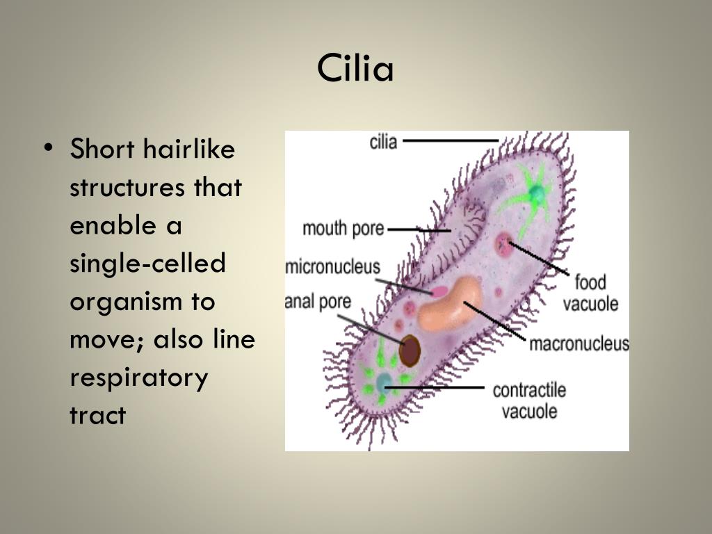
ciliate, any member of the protozoan phylum Ciliophora, of which there are some 8,000 species; ciliates are generally considered the most evolved and complex of protozoans. Ciliates are single-celled organisms that, at some stage in their life cycle, possess cilia, short hairlike organelles used for locomotion and food gathering.. The cilia are usually arranged in rows, known as kineties, on.
PPT Cell Organelles and Their Functions PowerPoint Presentation, free download ID2465516
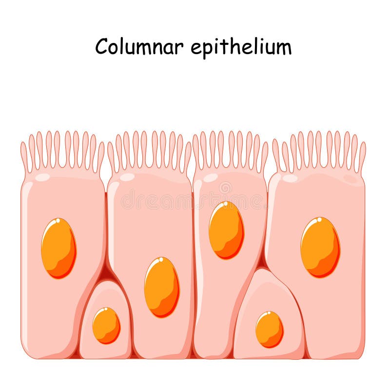
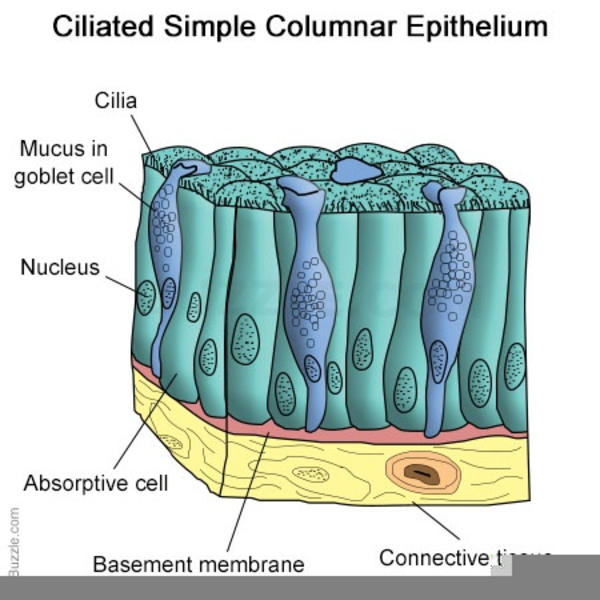
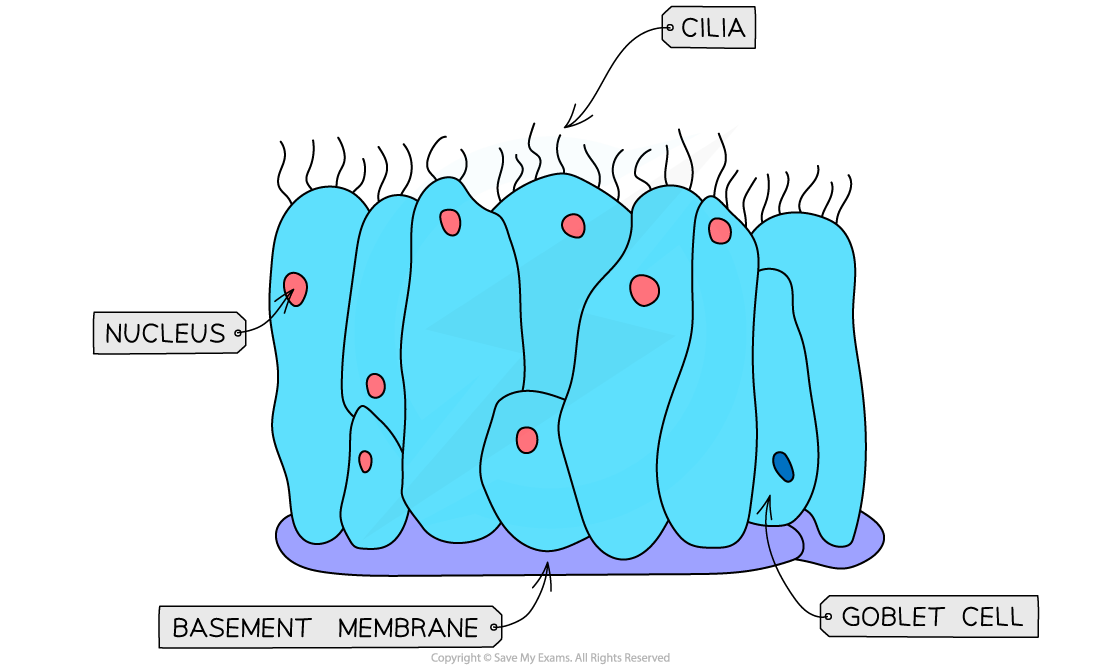
They are typically where absorption, secretion and filtration occur. The thinness of the epithelial barrier facilitates these processes. Simple epithelial tissues are generally classified by the shape of their cells. The four major classes of simple epithelium are: 1) simple squamous; 2) simple cuboidal; 3) simple columnar; and 4) pseudostratified.

Cilia & Mucus (11.1.2) CIE IGCSE Biology Revision Notes 2022 Save My Exams
The first pages illustrate introductory concepts for those new to microscopy as well as definitions of commonly used histology terms. The drawings of histology images were originally designed to complement the histology component of the first year Medical course run prior to 2004. They are sketches from selected slides used in class from the.

Structure of the primary cilium. The overall architecture and key... Download Scientific Diagram
Ciliated cells are located, with a small portion of cytoplasm, on the basement membrane and reach through the basal cells to the epithelial surface. The oval nucleus is present in the middle third of these cells. In the cytoplasm, which has a loose structure, scattered ribosomes are found, sometimes in groups.

Coloured TEM of a ciliated cell on human Stock Image P580/0099 Science Photo Library
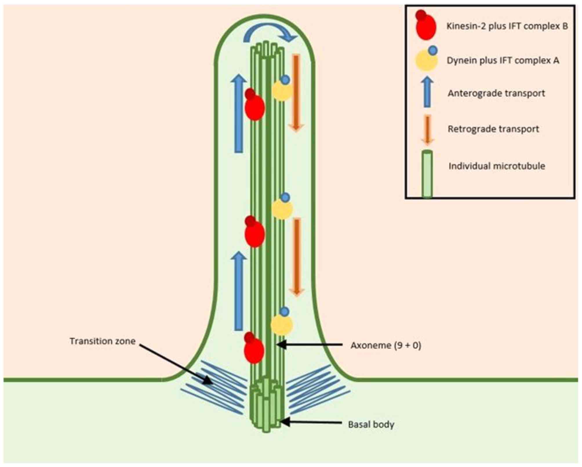
The cilium (plural: cilia) is a microtubule-based organelle that projects from the cellular membrane of many cells.Cilia can be divided into two types: motile and non-motile. Motile cilia sway in a wave-like motion in order to generate fluid movement. These types of cilia are found on the surface of cells such as the epithelial cells of upper respiratory and reproductive tract.
ciliated cell Labelled diagram
In multiciliated cells, the cilia become physically entrained to beat in metachronal waves in protists as well as in metazoans. This ciliated organism is a model of choice for the use of multidisciplinary approaches to dissect the location, structure and function of ciliary ion channels and other proteins involved in ciliary beating.
Ciliated Columnar Epithelium Stock Vector Illustration of education, lining 180548741
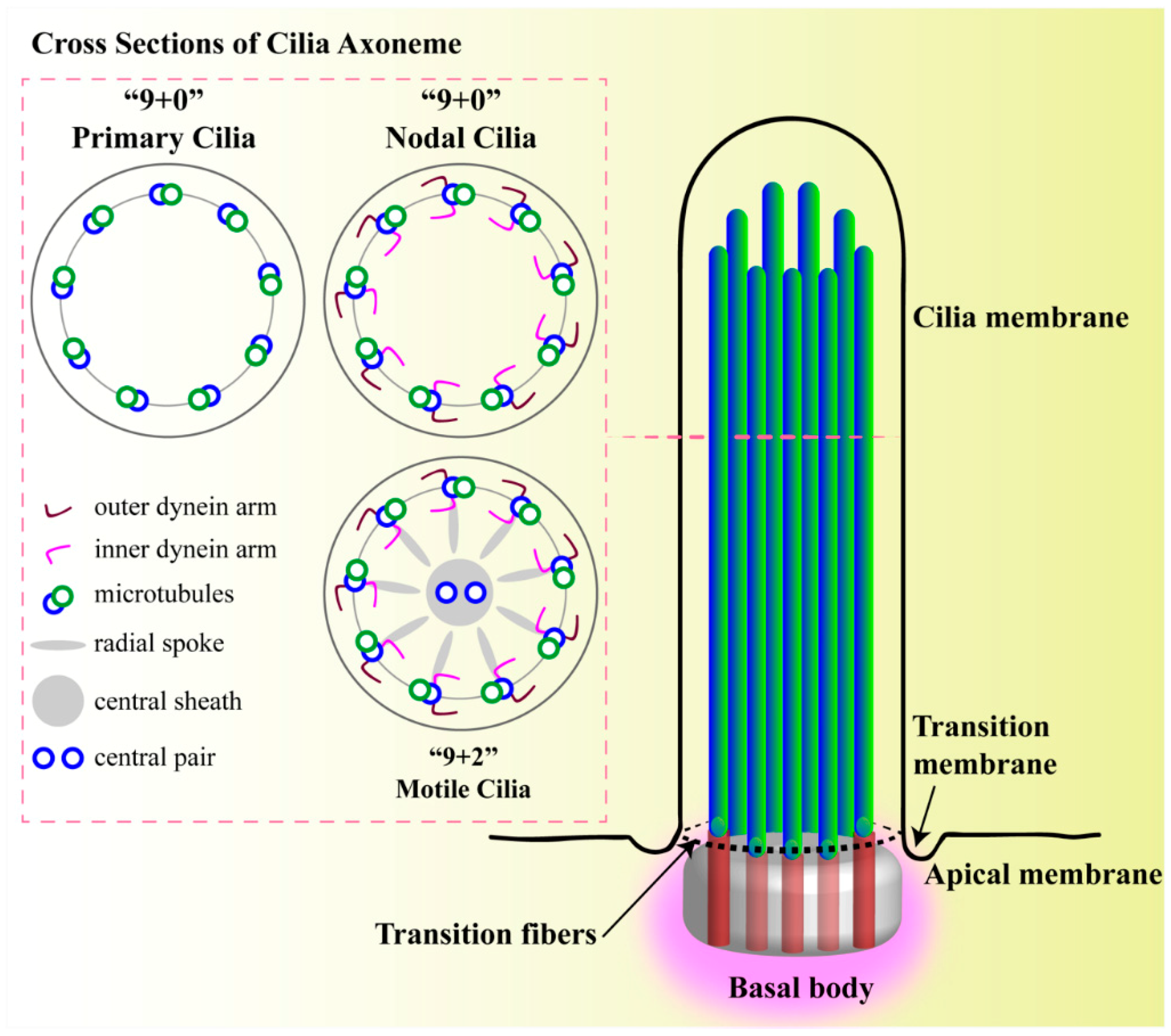
respiratory epithelial cells. (b) Structure of a 9 + 2 axoneme obtained by cryo-electron tomography. Density maps from subtomogram averaging (Bui et al. 2012) were combined. Important parts are. infect ciliated epithelial cells preferentially (Sims et al. 2008), and thus ciliated cell culturesareusedinCovid-19research.
Cilia structure Science, Biology ShowMe
Columnar ciliated cells are found throughout the. E. & Mukherjee, A. B. Uteroglobin: structure, molecular biology, and new perspectives on its function as a phospholipase A2 inhibitor..
squeamies — Ciliated Epithelium… Hairy Cells. Cells in your...
Cardiac (heart) muscle cells contract and relax to pump blood around our bodies for our entire lives. They never get tired. Smooth muscle cells make up thin sheets of muscle, such as the stomach.
Primary cilia and their role in cancer (Review)
The cell then divides in two, and each new cell obtains a copy of the micronucleus and the macronucleus. Ciliate undergoing the last processes of binary fission. Typically, the cell is divided transversally, with the anterior half of the ciliate (the proter) forming one new organism, and the posterior half (the opisthe) forming another. However.

Protista cillia flagella Cell theory, Nuclear membrane, Plasma membrane
Cells co-expressing gene markers of ciliated cells, including FOXJ1, and goblet cell genes such as MUC5AC, were termed 'mucous ciliated cells' and are postulated to represent a novel.

Cilia
Ciliated epithelial cells. In this clip the structure and function of a ciliated epithelial cell is described. Cilia are tiny hair like structures on the surface of the cell. The hairs sweep hair.
Ciliated Epithelium Structure Free Images at vector clip art online, royalty free
Ciliated Epithelium Cell - Concept. A section of epithelium is made up of columnar or cuboidal cells with hairlike appendages that can beat rapidly (see cilium). In structures like the trachea, bronchial tubes, and nasal cavities, the ciliated epithelium is responsible for transporting particles or fluid over the epithelial surface.
CIE A Level Biology复习笔记9.1.2 Distribution of Tissues翰林国际教育
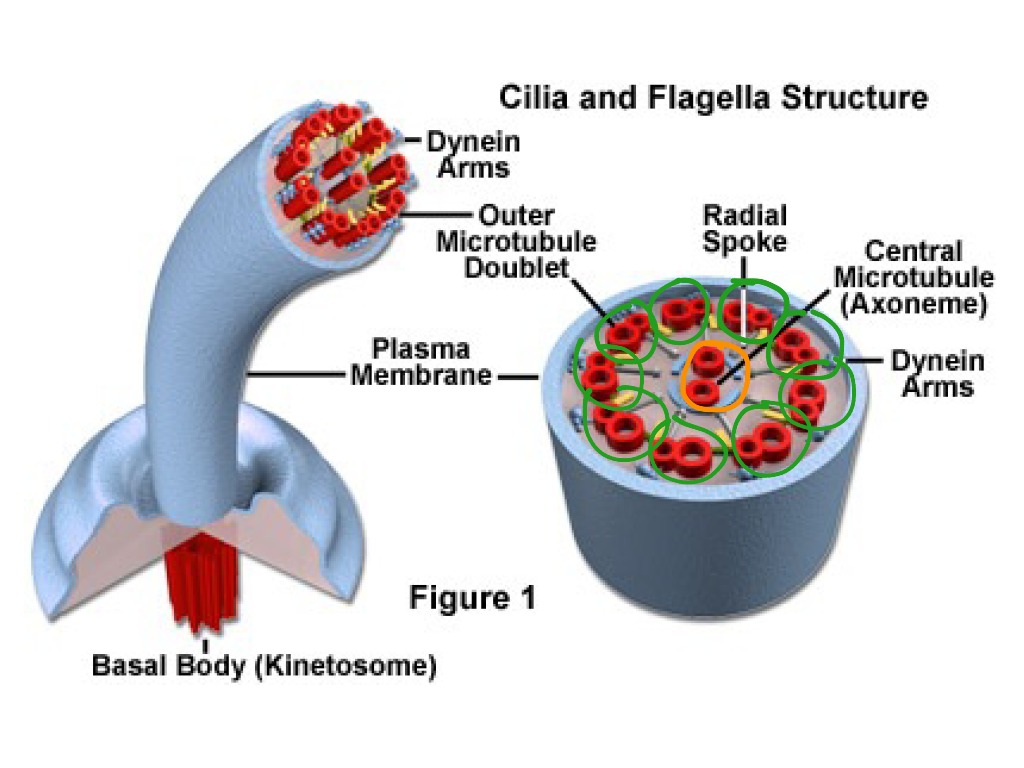
This structure is known as an axoneme, and the arrangement as '9+2', an arrangement ubiquitous in motile cilia. The microtubules are held together by cross-linking proteins. Between the nine outer pairs are motor proteins called dynein.. These ciliated epithelial cells remove mucus, bacteria, and other debris from the lungs. Another.
/ciliated_epithelial_cells-5a7cb8926edd650036eb92da.jpg)
Cilia and Flagella Function
5.6: Flagella and Cilia. Flagella (singular = flagellum) are long, hair-like structures that extend from the plasma membrane and are used to move an entire cell, (for example, sperm, Euglena ). When present, the cell has just one flagellum or a few flagella. Prokaryotes sometimes have flagella, but they are structurally very different from.

Cilia in the CNS The Quiet Organelle Claims Center Stage Neuron
The ciliated simple columnar epithelium bears cilia (finger-like projections of the plasma membrane) on the apical surfaces of cells, with goblet cells usually scattered throughout the epithelium. Up to 300 cilia may be present on each cell, beating in a synchronized manner with adjacent cells to propel materials over the surface of the epithelium.